The macules
Macules are flat, colored skin lesions that differ from the surrounding skin. They can come in a variety of colors, including red, brown, white, or other shades. The macules are not raised and usually do not cause changes in the texture or relief of the skin.
Macules can be caused by a variety of medical conditions, such as allergic reactions, infections, vascular disorders, pigmentary abnormalities, inflammatory reactions, and many more. They can be temporary or permanent, depending on the underlying cause.
Pityriasis versicolor
A superficial fungal infection of the skin that causes discolored, usually brown or light, spots on the skin.
Pityriasis versicolor, also called versicolor pitiriasis or pityriasis tinea, is an unsightly and non-contagious benign skin fungus of the skin caused primarily by a commensal yeast: Malassezia furfur (formerly known as Pityrosporum ovale).
This infection can cause discolored spots on the skin, usually in shades of brown, beige, or white.


Typical symptoms of pityriasis versicolor include:
- Round or oval spots on the skin, usually on the trunk, shoulders, neck, and arms. Only the palms and soles of the feet are never affected.
- The boundaries are clear and confluent, with trunk involvement.
- The spots can be of different colors, ranging from light brown to dark brown or white (depigmented forms exist.)
- The skin around the spots may be darker or lighter than normal skin.
- The spots may be slightly flaky, but they are usually not itchy or painful.
Pityriasis versicolor is more common in warm, humid climates. It can be aggravated by factors such as heat, humidity, excessive sweating, and hormonal imbalances.
The diagnosis is clinical or with the scotch test (with microscopic analysis)
Treatment for pityriasis versicolor usually involves the application of topical antifungal medications, such as creams, lotions, or shampoos, to the affected areas of the skin. In more severe or recurrent cases, oral treatment may be prescribed. It is important to follow your healthcare professional’s recommendations for the appropriate treatment
Fixed pigmented erythema
An allergic skin reaction to certain medications or substances, causing red and brown spots to appear on the skin.
Fixed erythema pigmentosa, also known as pigmented contact dermatitis, is an allergic skin reaction specific to certain substances. It does not necessarily cause red and brown spots to appear on the skin, but rather red, scaly lesions.
Common triggers for fixed pigmented erythema include certain medications, foods, chemicals, and cosmetics. Allergic reactions can vary in severity and duration.
Here’s how fixed pigmented erythema usually manifests itself:
- Onset of lesions : After initial contact with the triggering substance, red, swollen, itchy lesions appear on the skin. These lesions can be of different sizes and shapes.
- Brown colour : After the red lesions have disappeared, aBrown or pigmented oloration may remain on the skin where the reaction occurred. This is where the term “erythema pigmentosa” comes from. However, this brown coloration is often the next phase of the reaction and does not occur immediately after the appearance of the red lesions.
- Lesions recur : Lesions may recur in the same location if the skin is exposed to the allergen again.
The diagnosis of erythema pigmentata fixe is usually based on the symptoms observed and the patient’s history. It can be confirmed by skin allergy tests, such as patch tests.
Treatment of erythema pigmentata fixe involves avoiding the triggering substance, if identified. Antihistamine medications can be used to relieve symptoms. In severe cases, a doctor may prescribe topical corticosteroids or immunomodulatory creams to reduce inflammation.
“Café au lait” stains
Light to dark brown skin spots, usually present from birth. They are often harmless, but can sometimes be associated with genetic conditions such as neurofibromatosis.
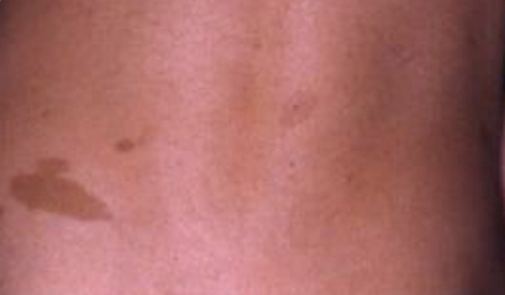
These are light to dark brown skin lesions that can be present from birth or appear soon after. Here are some more details about them:
- Appearance : “Café au lait” stains usually have well-defined edges and can vary in size. They can be small or grow to a larger size. The color can also vary, ranging from a pale brown to a darker brown, hence the name that evokes the hue of latte.
- Safety : In general, “café au lait” stains are harmless and usually do not cause symptoms. They are not painful and do not usually lead to medical complications.
- Genetic associations : Although most “café au lait” spots are benign, they can sometimes be associated with certain genetic conditions, including neurofibromatosis type 1 (NF1), also known as von Recklinghausen disease. Individuals with NF1 may have several “café au lait” spots, as well as other symptoms such as skin nodules, neurofibromas, and neurological problems. However, not everyone with “café au lait” spots necessarily has neurofibromatosis.
- Diagnosis and follow-up : The diagnosis of “café au lait” spots is usually clinical, based on the characteristic appearance of the lesions. If multiple “café au lait” spots are present or are associated with other symptoms, a doctor may recommend further evaluations to rule out underlying conditions such as neurofibromatosis.
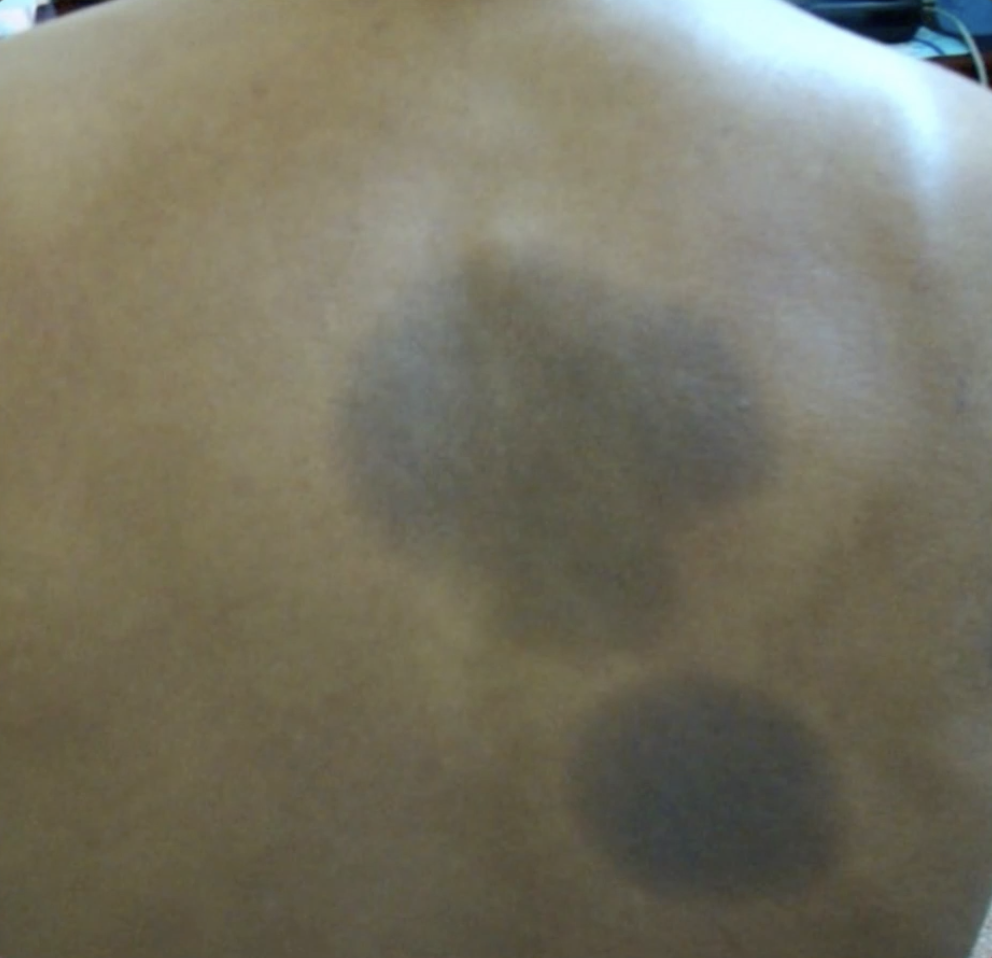
Mongolian Spot
A blue-gray or blue-black spot usually present at birth on the skin of some infants, more frequently seen in babies of Asian, African, Mediterranean, or Native American descent.
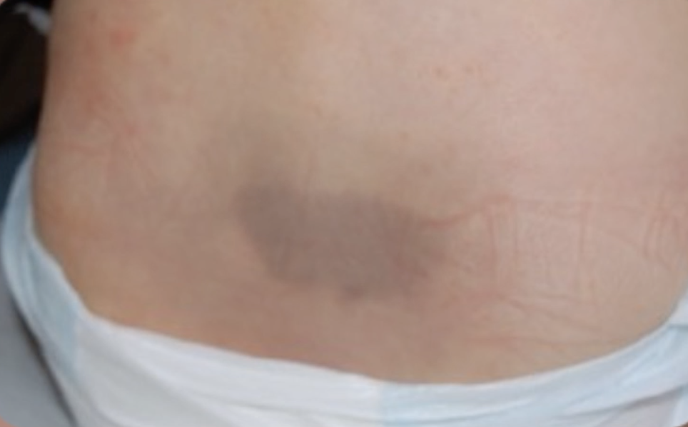
- Appearance : Mongolian spots are blue-gray or blue-black colored spots that are usually present at birth or appear shortly after birth. They are often oval or rounded and can vary in size, ranging from a few centimeters to several centimeters in diameter.
- Location : Mongolian spots are usually found in the lumbar region or on the buttocks, although they can sometimes appear on other parts of the body. They result from an accumulation of melanocytes (cells responsible for pigmentation) in the deeper layers of the skin.
- Ethnicity : Mongolian spots are more common in babies of Asian, African, Mediterranean, or Native American descent. They are less common in infants of Caucasian origin.
- Harmlessness : Mongolian spots are generally harmless and usually disappear on their own over time, usually in the first few years of life. They usually do not require any medical treatment.
It is important to note that Mongolian spots are a normal and benign feature in many infants and young children.
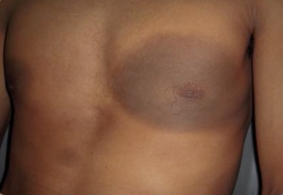
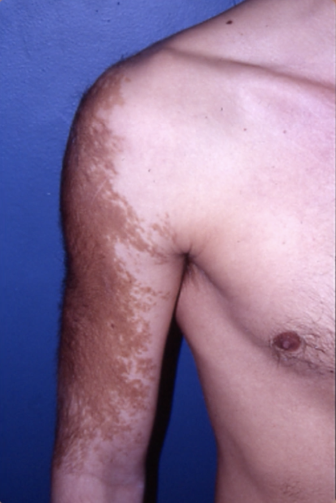
Becker’s nevus (or Becker’s hamartoma)
A rare skin lesion that usually develops in adolescence, causing a pigmented, irregular skin patch.
- Appearance : Becker’s hamartoma is a benign skin lesion that usually presents as a pigmented, irregular patch of skin. This spot can vary in size, shape, and color. It can be brown, reddish, or other pigmented hues.
- Development : Becker’s hamartoma tends to develop during adolescence, although it can also appear later in life. Boys are more often affected than girls. The lesion may increase in size gradually over time.
- Location : Becker’s hamartoma lesions can develop on different parts of the body, including the shoulders, trunk, arms, legs, and sometimes the face. They can be single or multiple.
- Safety : In general, Becker’s hamartomas are mild and do not usually cause serious symptoms or health problems. However, they can sometimes be associated with aesthetic discomfort for some people, especially because of their appearance.
- Treatment : Most of the time, Becker’s hamartomas do not require medical treatment. However, if the injury causes physical or emotional discomfort, treatment options such as laser therapy, cryotherapy (freezing), or surgery may be considered to reduce the appearance of the injury
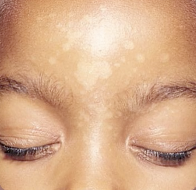
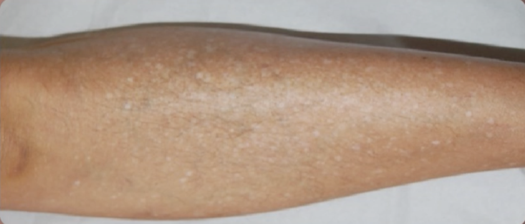
Idiopathic leukoderma (hypomelanosis)
A skin condition characterized by white or depigmented spots of varying size, often seen after skin inflammation or skin aging (helioderma) related to prolonged sun exposure
Idiopathic hypomelanosis is a skin condition characterized by the appearance of small, depigmented spots on the skin. It is often also referred to as “pityriasis alba”, although this term is sometimes used to describe other similar conditions. Drop idiopathic hypomelanosis spots are usually lighter in color than the surrounding skin and may look like drops.
Here are some characteristics and details about Idiopathic Hypomelanosis Drops:
- Appearance : Depigmented spots associated with Idiopathic Hypomelanosis Drops are usually small, round, or oval, and can vary in size. They are often white or very light in color, making them distinct from normal skin.
- Location : Spots can appear on different parts of the body, but they are most commonly seen on the arms, legs, shoulders, face, décolleté and neck.
- Cause : The term “idiopathic” means that the exact cause of this condition is not fully understood. However, factors such as sun exposure, a mild inflammatory reaction, or pigmentary imbalances may play a role in its development.
- Safety : Idiopathic hypomelanosis is usually mild and does not usually cause symptoms other than the appearance of spots.
- Treatment : Typically, treatment is not necessary, as the condition tends to improve on its own over time. Using moisturizers and proper sun protection can help reduce the appearance of dark spots.
Leprosy
An infectious disease caused by the bacterium Mycobacterium leprae, which can cause hypopigmented or depigmented skin spots in people with lepromatous forms of the disease.

- Transmission : Leprosy is mainly transmitted through prolonged and close contact with an infected person. However, it is important to note that leprosy is relatively non-contagious and the vast majority of individuals are naturally immune to the disease.
- Symptoms : The symptoms of leprosy vary depending on the form of the disease. In milder forms, called “pauci-bacillary”, symptoms can be mild and manifest mainly as hypopigmented or reddish skin spots, numbness or anesthesia in the skin area and tingling. In more severe forms, called “lepromatous,” the disease can cause larger skin lesions, nodules, deformities, nerve damage, and other complications.
- Treatment : Leprosy can be effectively treated with antibiotics such as rifampicin, dapsone, and clofazimine. Early treatment can help prevent complications and limit the damage caused by the disease. Treatment is usually given over an extended period of time and under the supervision of a healthcare professional.